Asbestos in Spray-on Fireproofing Containing Vermiculite (SOF-V) Webinar Transcripts
Webinar Series Transcripts

Webinar #1 – Amphibole Asbestos in Vermiculite

Webinar #2 – Asbestos Detection in SOF-V LAB.055.1 Method Overview

Webinar #3 – Asbestos Detection in Vermiculite Spray-on Fireproofing (SOF-V) – Item No. 198.8


Webinar #1 – Amphibole Asbestos in Vermiculite

Click here to download the webinar PDF

Presenter:           Matthew S. Sanchez, Ph.D.

Date:                    October 29, 2014

Slide 2
Narrator: Changing regulations and requirements make it challenging to stay current, so we put together this webinar to clarify NY State’s guidelines and to explain the techniques available for asbestos analysis. RJ Lee Group has pioneered the development of asbestos analyses methodologies for more than 20 years.

Before 1990, some vermiculite used in building materials was found to contain asbestos, especially in spray-on fireproofing or SOF-V.

Regulated and non-regulated amphiboles meet the Environmental Protection Agency’s definition of asbestos. Across the U.S. many buildings still contain asbestos or SOF-V that could contain asbestos. The NY State Department of Health determined existing methods of analysis were insufficient to quantify low levels of asbestos, and that SOF-V containing more than 10% vermiculite must be considered asbestos-containing material or ACM. Two methods are currently accredited for asbestos analysis in NY State – 198.8 and RJ Lee Group’s Method LAB.055.1. Our experts can now quantify regulated and non-regulated more accurately than ever before.

I’d like to introduce our speaker today, Dr. Matthew Sanchez. Dr. Sanchez has been involved in the characterization of vermiculite and asbestos-associated amphiboles for more than a decade. The research from both his masters and doctorate degrees are directly related to both the geologic and environmental issues surrounding the former vermiculite mine in Libby, Montana. He has also been involved in both method development and analysis of asbestos in air, bulk materials, soil, water, and industrial minerals. Dr. Sanchez.

Dr. Matthew Sanchez – Presentation

Hi, I’d like to welcome you all and thank you again for giving us some time today as we go through some of the vermiculite work going on in New York. We’ll talk about some of the analytical solutions for the choices you have before you.

Slide 3
We’re going to go through a little bit of the timeline: the DOH (NY Department of Health) letters and FAQ #10 regarding vermiculite. The methods that have been approved to date are only for those thermal insulation systems that can contain the vermiculite spray-on fireproofing. We’re going to go through and compare directly our method LAB.055.1 with the NY DOH 198.8 method.

The concern with vermiculite by the DOH was stated in a guidance letter they issued on June 22, 2012 to clarify one of their FAQs they wrote about vermiculite, and this quote is taken directly from that guidance letter – “where vermiculite is determined to be more than 10% in the material, that material must be reported as an asbestos-containing material by laboratories.”

Slide 4
Some further explanation in that same letter is this next quote (on the slide and added here) taken from the EPA’s website regarding vermiculite—“However, because vermiculite’s asbestos contamination typically ranges from 1% to 5%, vermiculite’s contribution to asbestos content of vermiculite materials used for thermal systems insulation, surfacing materials and other miscellaneous ACM . . .” This information directly relates to vermiculite from Libby, MT which was mined up until about 1990, and some of the stockpiles continued to be sold for few years thereafter.

And in that vermiculite, depending on different reports that you can find and read, these concentrations go from low quantities to potentially up to 5% in the raw materials. So, again, the assumption made here is that the vermiculite could be contaminated with asbestos coming from the Libby vermiculite mine.

The amphibole asbestos from Libby is different from that typically dealt with, in that the mineral is an amphibole-type asbestos and the predominate type is called winchite and,  by name, winchite is not a regulated mineral.

Slide 5
Let’s review what the regulated asbestos types are, and what the EPA and the NYDOH use in their regulations as well. You have one that belongs to a group of minerals called serpentine, but chrysotile and the rest of them come from the amphibole, bi-amphibole group. This is a group of minerals of different compositions but very similar structures.

These by name are amosite, crocidolite, anthophyllite asbestos, tremolite asbestos, and actinolite asbestos.

Slide 6
A few months after that initial guidance letter, another guidance letter came out with some more clarification. The main difference in this letter was that it now allowed those who had MSDS sheets and knowledge of where their material came from and documentation saying it was not ACM, to be sufficient. They wouldn’t have to go back and re-test it. They would assume it was ACM if it had greater than 10% vermiculite.

We were aware of these issues as a NY ELAP-certified lab, but we really got heavily involved in the vermiculite issue in January of 2013. Again, this was post-hurricane Sandy up there in New York and along the Jersey coast. We had a client approach us and they were looking for a lab that could work with them and be able to take a method to the State and be able to work with the State to resolve this issue that they were facing. They had floors that needed to be renovated from damage from the storm, and under these guidances that they were dealing with was saying they had to abate all this material. It was that kind of a situation. Our work with the State was very intensive and ongoing. I’ll describe some of the validation that we had to do in order to satisfy the state to show what our method was capable of doing. In our data validation packages, there were many deliverables on kind of a timeline – April 2013, June, July. There was a bigger hiatus there as we waited to hear back.  Then some final data was provided to NY DOH to satisfy their concerns in January 2014, and then the final one was in February after we actually ran a PT sample for them, or at least a test sample as they called it, since we were the only lab that got it at that time. This process has been going on for almost two years now.

Slide 7
In the middle of all that – some of these validations and things – while we working on that, they came out with another guidance letter. This was July 9, 2013. They started to allow the use of 198.6 as long as they had this disclaimer – the specific disclaimer “This method does not remove vermiculite and may underestimate the level of asbestos present in a sample containing greater than 10% vermiculite.” This was progress it seemed, but you still had this ambiguity—Is there asbestos there? Is there not? Kind of unsatisfactory for people in health and safety and for the interest of their clients.

Slide 8
The latest guidance on this issue came out just a few months ago, on July 22, 2014. This, again, is taken directly from them. They announced “. . . the imminent availability of two new NYS DOH ELAP-approved methods for the detection and quantitation of asbestos content in spray-on fireproofing that contains vermiculite (SOF-V).” One of these methods is the RJ Lee Group Method LAB.055.1. The other method is promulgated by the State, and it was developed in conjunction with Dr. Chatfield and a lot of support from REBNY (Real Estate Board of New York) up there in New York, and it was titled 198.8. Also, in that letter, they stated that two days from now on October 31, 2014, one of these two methods must be used on these SOF-V materials.

Slide 9
Since July 25 (2014), our lab has been accredited through the NY State ELAP program to run our method and be accepted in the state for these vermiculite-containing groupings.

Slide 10
A little more on the guidance, it goes into another section on industrial applications on how these methods need to be employed moving forward. I’ll briefly go through these, but I recommend you read through yourself to understand some of these responsibilities.

  • For all future projects with no associated surveys, you must go and re-sample and test using one of these two methods.
  • Projects with existing surveys but no project plan, you need to get back in there and re-sample and re-survey, unfortunately.
  • Projects with existing surveys and projects yet to be done, known as the Phase 1A period, must re-sample and re-survey.
  • Projects in progress using an old survey with work underway: they’re recommending you go in and re-sample, but they’re not requiring you to unless there’s some kind of a change order on the work being performed.
  • For projects that have been completed under old surveys, those are just fine as-is. However, if those materials need to be disturbed or worked on again, a new survey is going to need to be done for those.

Slide 11
We’ll move on now and try to contrast the two methods available as options. Again, these are the RJ Lee Group Method and the NY 198.8. I’ll be highlighting the differences between the methods. I’ll try to give you a better idea of what your options will be.

In our method (LAB055.1), we actually dissolve the vermiculite matrix, so we get rid of the vermiculite that is there. The concept behind this is very similar to what you may be familiar with as the NOB samples or the Non-Friable Organic Bound analyses. Our method uses both PLM and SEM — SEM is similar to TEM in that it’s electron microscopy and allows a little higher magnification. The benefit it gives in this analysis is that it allows us to obtain some compositional data comparable to the EDS on the TEM. This allows us to differentiate the non-regulated species of amphibole asbestos from regulated amphibole asbestos types. Again, this is very important when you are dealing with these materials that are potentially coming from the Libby, MT source. The composition of those amphiboles, you know, is not tremolite, not actinolite, they’re these winchite series amphiboles.

The NY method (198.8) uses a density-separation method using some heavy liquids. They’re using different densities of the amphibole minerals with the vermiculite and other materials to be able to separate those out. A crude analogy would be like panning for gold, looking for the heavy gold is some kind of lighter sediment. Their method uses only PLM for the identification and the quantitation of the asbestos. This is the big difference between the two methods. Using only PLM, you’re not going to be able to tell apart the tremolite from a winchite based on the optical properties. I’ll show more information about that here in a few more slides.

Slide 12
Other big differences with the methods is going to be how these methods have been validated and the labs that are performing them, what’s been required of them (the labs) to be able to do the analysis. Our validation work with NY State was very extensive. They required us to make standards of all six regulated types of asbestos, different concentrations. Based on some of those results, we went back in and created more standards closer to that 1%, or the ACM criteria, to really test the method and see how well it performed. The validation that NY State did was performed internally. Ours was actually presented at an ASTM meeting this past July in Vermont, where they used one type of asbestos in different concentrations in their validation. The way this works is that labs that seek the accreditation have to provide certain documentation. One of the things they need to do is to provide a DOC, or a Demonstration of Capability, for the analyst who will perform the method. The minimum requirement for this DOC documentation is four samples, and it can actually be the same sample just analyzed four times.

Slide 13
To speak more about what we did internally here, working with the DOH, we ended up having four separate analysts perform triplicate analyses. There were many, many data points that were collected. We also had experience up to this point with our method analyzing several hundred commercial tests, real products that were coming out of buildings at this time. Our samples were composed of 5-gram base samples, and again, we spiked these with known concentrations of the different regulated asbestos types. What we needed to prove to DOH was that our method was as well or better than the existing methods.

Slide 14
So we actually compared our data to both 198.1 and 198.6, with 198.6 performing much better relative to 198.1. The next slide is just a portion of this validation data dealing with detection of the amphibole-type asbestos using our method and comparing it to the 198.6 method. On the vertical scale on this graph is the observed and measured concentration reported by running the two methods. The horizontal scale is the known concentration – how much we knew we put into these samples. As you look at the concentration, the black dots are the RJ Lee Method and the red is the NY 198.6 method. The RJ Lee Group method is much tighter in its distribution of reported percentages for a known concentration. As you start looking down here at the 0.5 and the lower concentrations, these concentrations represent non-ACM materials. These are less than 1% by weight. What you find is that, in our method, when you start finding these low levels of 0.5 and .25% amphibole levels, our method is nearly 100% accurate in our recoveries of the amphibole added. When running the 198.6, in the case of the 0.5, almost all the analyses reported out as a false positive. These would have been reported out as a positive material when, in fact, they were negative.

Slide 15
To elaborate a little more on the method and what these things would look like, this picture on the left is an SEM image of a filter residue. This is after our method preparation has been performed. Approximately 98% of these materials get dissolved in the ground fireproofing, and this is what is left behind. This is a sample that was spiked with .1% amphibole from Libby, MT as an example of what these samples would look like. The highlighted, kind of elongated particles in red, are amphibole asbestos particles. There are many more there. These are just shown to illustrate what you should be looking at here. The smaller image off to the right shows what the composition of that asbestos fiber is. That composition containing those different elements that are labeled Na, represent sodium and so forth. That’s the composition of that fiber. You can then further identify what type of amphibole that is. That kind of composition is characteristic of material from Libby, MT. It’s a winchite amphibole.

Slide 16
This is what a positive sample at the threshold of 1% would look like on our residues. So, between these two slides, you go from, “yeah there’s some there,” to almost everything you see is potentially amphibole asbestos. So the determination of these things as to whether they are asbestos-containing or not becomes very apparent when you start getting high enough concentrations to get you over that 1% ACM level.

Slide 17
Let’s go a little further. Those slides were from the SEM analysis. Let’s go into some of the PLM analysis work. The image on the left is a photograph of the filter, and the circled particle is a bundle of amphibole asbestos that you can readily pick off. The picked-off particles are on the right and you can measure the optical properties and quantitate this is in regard to the residue remaining. This is a real-world sample. These are the kinds of concentrations typically we’re dealing with.

Slide 18
Here’s a similar real-world sample (on the left): an SEM image, showing the amphibole asbestos once the vermiculite and other materials have been digested away. On the right is another example of the composition of those particles, and again, we’re looking at a Libby source material—winchite.

Slide 19
Moving on and speaking to 198.8, we look forward to hearing from the NY DOH internal labs that performed this work for validation. They did say in July that they will be publishing findings. We look forward to seeing those so we can do some further comparisons of our method to 198.8. Initially, we ran 198.8 in our laboratory and we’ve also applied for accreditation for that method, as well. There are two analytical problems associated with 198.8 on the first pass through.

  • The first I already mentioned, and this is dealing with the inability of PLM to differentiate between the asbestos types that are lower in vermiculite than those that came from Libby, and what the regulated forms are. This, again, is speaking to the winchite, tremolite/actinolite differentiation.
  • The second is the potential for the amphibole asbestos to become trapped in the vermiculite float.

We’ll highlight both these points in the next few slides.

Slide 20
When you are doing the PLM analysis, when you go in to identify the asbestos types present, you’re measuring different properties using the light microscope. One of the most important properties that you can measure to tell whether this material is amosite or crocidolite or tremolite or chrysotile, is dealing with the refractive indices. With these regulated amphiboles, the refractive indices are different enough for most of them so that you can readily distinguish them simply by that measurement. So, in this graph, from a publication from McCrone, on the very top line where it is giving the different refractive indices values for tremolite, it is showing a kind of range that is observed in nature for these materials. The lowest row labeled “Thesis Index Range” is taken from a thesis by Bryan Bandli. I think it was published in 2003. In the thesis, they did precise optical measurements of the refractive indices and material specific to the Libby vermiculite and the amphiboles that occur in that vermiculite deposit. The take-home here is that, if you compare these ranges that you would expect tremolite to fall within and expect the winchites from Libby to fall in, they overlap. So, based on what you can measure optically, you cannot tell these two minerals apart – these two amphibole species apart. Again, this was adapted from a paper by both Millete and Bandli in 2005.

Slide 21
To go on about potentially losing some of the amphibole in the float, according to 198.8, you actually discard the float after your heavy liquid separation. So, theoretically, in the separation, the amphiboles will drop out during the centrifugation and the vermiculite will then be floating and then it’s just a matter of decanting off the vermiculite and recovering the amphiboles. So, theoretically, this works well based upon the density. However, the nature of how the amphibole occurs in these materials, especially from the Libby source, is that the amphibole actually forms in place with the vermiculite such that the amphibole asbestos can be bound between the sheets of the vermiculite. There is also the potential for these amphibole fibers to be somewhat adhered, or to ride the vermiculite that floats up. They’re not getting fully in contact with these heavy liquids and thus be able to drop out. As an example of that, on the samples we ran for our Demonstration of Capability, instead of throwing away the floats, we actually went through and looked at them on the PLM. In fact, you can occasionally find amphibole asbestos in the float material. So, there’s a potential there of losing some of the asbestos during your preparation.

Slide 22
I just want to summarize where we are to date. One year ago we were in a situation where labs pretty much had to report out these things as ACM or, at best, they could put a disclaimer. Today there are two methods that have been approved for use in the state of New York: one is our method LAB.055.1 and the other is the NY DOH method 198.8. Some of the highlights that make our method very good and very useful is that it is able to discriminate the types of amphiboles and, without that floating step, we’re dissolving everything and putting it down on the filter. The chance of losing these materials, losing asbestos at these low levels, becomes very, very minor. It reduces that potential for error.

Slide 23
Ultimately, we worked really hard on this. We worked a lot with the NY DOH. There were very many back-and-forths, more data generated. Looking at the data, it is a good example of industry and regulatory body cooperation in trying to solve a problem. This also includes the work done by Dr. Chatfield and the NYS DOH internal lab to make these options available to resolve this issue.

We’ll be wrapping this up and thank you for your attendance today.


Webinar #2:   Asbestos Detection in SOF-V LAB.055.1 Method Overview

Click here to download the webinar PDF

Presenter:          Craig Huntington

Date:                  November 19, 2014

Moderator:    
Slide 1
Good afternoon everyone, and welcome to Part 2 of our webinar series on the regulation of spray-on fireproofing containing vermiculite brought to you by RJ Lee Group. I’m Kathy Gilkey and I will be the moderator for today’s event. I’d like to take a few moments to go over some information to help the presentation run smoothly. The presentation will last approximately 20 minutes. Following the presentation we’ll have a short question and answer session. If you have a question, please type it into the chat box on the lower right of your control panel. Only the moderator will see your questions. We will answer as many questions as possible during the question and answer session. We’ll answer any remaining questions off-line and provide the answers to you directly.

Slide 2
Previously we covered topics related to the historical perspective on vermiculite and vermiculite regulations. We also briefly discussed the two approved methods for SOF-V analysis, as well as some details surrounding the validation of methods. Today we will dive deeper into RJ Lee Group’s LAB.055.1 Method. If we go back even as recently as a year, materials containing over 10% vermiculite were considered ACM (asbestos-containing material).

Slide 3
Now we have two methods available to alleviate having to make that assumption. Since we are past the October 31 deadline, one of these two methods must be used for the detection and quantitation of asbestos content in SOF-V: RJ Lee Group’s LAB.055.1, which we are covering today, or the ELAP method 198.8. LAB.055.1 is designed to detect and quantitate all six regulated types of asbestos, but also differentiate them from any non-regulated types of asbestos contained in the sample. Today we present a LAB.055.1 method overview: determination of asbestos in spray-on fireproofing containing vermiculite (SOF-V).

Slide 4
In July 2014, the NY State DOH announced the approval of two methods to determine asbestos in spray-on fireproofing containing vermiculite. RJ Lee Group was the first to receive approval for their method LAB.055.1. This analysis avoids the false positives that may come from the presence of non-regulated asbestos, and the false negatives caused by asbestos trapped within the vermiculite that can’t be measured. Today, my colleague, Craig Huntington, will discuss LAB.055.1 in more detail. Craig has over 30 years’ experience as a geologist. As a materials specialist and consultant for RJ Lee Group, he has worked on several high-profile projects related to asbestos including developing a portable elutriator for measuring re-suspended surface dust at a building affected by the World Trade Center disaster. Now, I’d like to introduce, Mr. Craig Huntington.

Craig Huntington – Presentation:

Slide 5
Thank you, Kathy. We’ll begin with the introduction of the RJ Lee Group LAB.055.1 method for the analysis of asbestos in spray-on fireproofing (SOF-V).

Slide 6
The purpose of the analytical method is to determine the total asbestos content in the six regulated asbestos minerals in vermiculite spray-on fireproofing.

Slide 7
The process involves adjusting large amounts of the cellulose, gypsum, vermiculite in carbonates, generally anything that is combustible or readily soluble, in order to detect asbestos and lower the detection limit.

Slide 8
In our macro examination, when the sample arrives at the lab, we first determine whether the sample contains layers. If so, each layer is treated as a separate sample. Then we check to see if the sample contains vermiculite. If so, it is readily applicable to the LAB.055.1 method.

Slide 9
As an example of how the digestion of the sample affects the appearance in the analysis, here is a process overview of the LAB.055.1 method. It consists of two levels of analysis: Level I is for the analysis of chrysotile in the sample by PLM (polarized light microscopy). Level II is for the analysis of asbestiform amphibole in the sample by PLM and SEM (scanning electron microscopy). Both levels of analysis reduce the matrix by ashing at low temperature and by digestion.

Slide 10
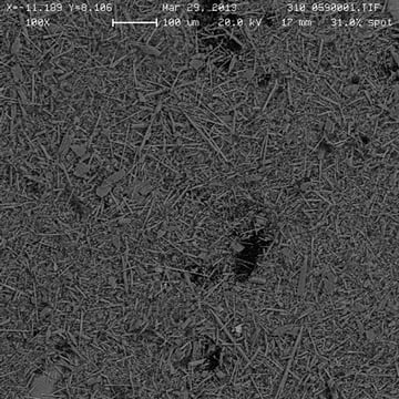
This photo gives an example of how the digestion of the sample affects the appearance in the analysis. In the left image, we have raw spray-on fireproofing. It is very difficult to identify either chrysotile or any of the regulated amph ibole asbestos minerals in this type of unprocessed material. On the right, we have an SEM image of Level II-processed samples. In this case, the matrix has been reduced by about 98%. So we’re looking at a 2% residue in the sample and we can readily pick out in this image asbestiform amphibole bundles, shown by the yellow circles. By removing the matrix material, the asbestiform amphiboles are much easier to observe, measure, and quantify.

Slide 11
The LAB.055.1 begins at Level I treatment for chrysotile analysis with a 400-point count. If there’s less than ½ % of chrysotile, we proceed immediately to Level II. If there’s greater than 2% chrysotile, we report the sample as ACM. If there’s between ½ and 2% chrysotile, then we repeat the analysis in triplicate and average the results to determine the concentration. If the mean of the three is less than 1%, we proceed to Level II. If the mean is greater than 1%, then we report the sample as an ACM material.

The Level II treatment is for the analysis of amphiboles. It utilizes both polarized light microscopy and SEM. If there’s less than 1% regulated amphibole asbestos, we sum the results for Level I for chrysotile and Level II for regulated amphibole asbestos, and if the mean is less than 1%, then we report the sample as non-ACM. If the mean is greater than 1%, we report the sample as SEM. If there’s more than 1% regulated amphibole asbestos in the sample, then we do report the sample as ACM.

Slide 12
Now, let’s look at a microscopic analysis of some of the materials that obscure the asbestos in the spray-on fireproofing.

Slide 13
One of the principal materials is vermiculite. Vermiculite is a clay mineral produced by low temperature alteration and weathering of mica. And as you can see from the formula, (Mg,Ca)0.7(Mg,Fe+3,Al)6.0[(Al,Si)8O20](OH)4 8H2O, it has eight molecules of structural water.

Slide 14
When heated rapidly up to 300 ˚C, this generates steam which expands the flat sheet structure exponentially and drives the layers apart. In the left-hand photograph, we see sheets of vermiculite, flat sheets with perfect basal cleavage. On the right, we see an expanded vermiculite particle in which the expansion can be up to 30 times. The material has very low density and can actually float on water.

Slide 15
Now we’ll look at some of the optical properties we use in polarized light microscopy to determine the nature of minerals. Vermiculite is a moderately pleochroic mineral. That is, it changes color from tan to brown when the mineral is rotated on the stage. It exhibits what we call moderate birefringence. Birefringence is really the difference between the highest and lowest refractive index of the mineral. It exhibits a positive sign of elongation along the cleavage traces. The sign of elongation is the relationship between the crystallographic axis and the principal vibration direction. As you can see from these images, vermiculite has perfect basal cleavage and it sits down flat on the surface of the slide. And, as shown on the left-hand photograph which is a plane polarized light photograph, it is somewhat opaque and can obscure other particles beneath it. This is the principal reason we’d like to get rid of this material in doing the analysis. That same particle is shown in the right photograph in cross-polarized light. This image shows the cleavage traces on the right-hand corner and here, these are the edges of the plates. They are not fibers in this particular image.

Slide 16
The next particle we are interested in is a common constituent in spray-on fireproofing and it is cellulose. Cellulose is paper fibers. This material is added to another material to help bind it together and it’s very low cost. As you can see in these images, it’s quite birefringent and it occurs typically as flat ribbons. So as not to confuse the cellulose fibers with asbestos, we remove the cellulose by ashing at low temperature.

Slide 17
Other fibers that we have added to spray-on fireproofing are chrysotile or fiberglass, based on the age of the material. Prior to the introduction of the asbestos regulations in the 1970s, chrysotile was added to the fireproofing to provide mechanical strength and spalling resistance. Chrysotile does not normally occur with vermiculite deposits, but only is added – and was added – in materials prior to the 1970s. After the asbestos regulations were introduced, fiberglass was substituted for chrysotile for mechanical strength and spalling resistance in lieu of chrysotile.

Slide 18
Chrysotile is an asbestiform serpentine mineral and was mined and used extensively in North America. As you can see here in the formula, it’s magnesium silicate and it occurs as a rolled sheet that forms a hollow fiber. The hollow-fiber nature of chrysotile gives it excellent insulation properties.

Slide 19
In polarized light microscopy, chrysotile is colorless. It occurs in fiber bundles with low birefringence and low relief in a 1.55 refractive index oil. It exhibits positive elongation and is asbestiform. The refractive indices are typically in the 1548 to 1556 range. On the left-hand photograph in plane-polarized light, we see the bundle extending horizontally, as shown by the arrow, and that same fiber bundle is shown on the right in cross-polarized light.

Slide 20
When we conduct Level I analyses, we remove the gypsum, the cellulose and the carbonate from the sample by low-temperature ashing and by washing with water and mild acid. This sample shows thin, tan vermiculite flakes and a large chrysotile bundle as pointed out by the arrows in the left-hand photograph. And, in the right-hand photograph, the chrysotile bundle, the blue bundle, is running underneath the vermiculite flake but you can still see it in the image on the right. For more than 35 years, fiberglass, rather than chrysotile, has been added to fireproofing.

Slide 21
Fiberglass is amorphous and has no crystal structure. The image on the left shows raw fireproofing with a bundle of fiberglass sticking out of it. Attached to the fiberglass fibers are bits of gypsum. Gypsum is the binder for this particular bit of spray-on fireproofing. In cross-polarized light, fiberglass is isotropic and it is only shown in the right-hand photograph by the presence of gypsum particles adhering to it.

Slide 22
Vermiculite spray-on fireproofing may also contain amphibole minerals which may be asbestiform, non-asbestiform or both. Asbestiform amphiboles found in fireproofing typically occur as individual bundles or are inter-grown with the vermiculite sheets; they are not added as a separate constituent to the fireproofing. The amphibole minerals associated with vermiculite deposits are typically anthophyllite, tremolite-actinolite or richterite-winchite.

Slide 23
To determine whether a particular amphibole particle is asbestiform or not, we rely on the EPA guidance in the form of EPA 600 which defines asbestiform morphology under the microscope as having the following characteristics, particles with:

Mean aspect ratio greater than 20:1 for fibers longer than 5 microns

  • Very thin fibrils, usually less than 0.5 microns in width and
  • Two or more of the following properties:
    • Parallel fibers occurring in bundles
    • Fiber bundles displaying splayed ends
    • Fibers in the form of thin needles
    • Matted masses of individual fibers, and,
    • Fibers showing curvature

Slide 24
This particular slide shows fiber bundles that were hand-separated from a sample of expanded vermiculite. In the upper, left-hand photograph, you can see bundles, rather large bundles actually, (because the scale bar there is 2 mm of amphibole asbestos) were picked out of a sample of expanded vermiculite. In the center slide, we’ve taken one of those bundles and crushed it down and you can see running horizontally across the slide, thin fiber bundles of amphibole asbestos. In the lower-right photograph in cross-polarized light, we can see that those fiber bundles in the center are near extinction when oriented in the east-west orientation parallel to the polarizer.

Slide 25
The asbestiform amphibole can also be an inter-growth with vermiculite. In this particular series of photos, in the upper left, we have a binocular photo that shows vermiculite flakes or mica flakes on the surface of the filter and there is a fan-shaped amphibole asbestos bundle identified by the arrow. That bundle was removed from the filter and then placed in immersion oil. In the center photograph we see that plane-polarized light and you can see the close proximity of the fan in association with the vermiculite plate or flake. And then in the lower right, we can see that same bundle in cross-polarized light.

Slide 26
The optical properties of the amphiboles that typically occur with vermiculite, that is anthophyllite, tremolite-actinolite, and richterite-winchite, have optical properties that are very similar and they tend to overlap. In the chart, going from left to right we have the lowest index for each of those mineral groups, followed by the higher index and gamma, and then the birefringence. You can see that these properties or values are all very close and overlapping. All these minerals exhibit positive elongation, and they are all pleochroic in different shades of pale brown and green. The pleochroism itself is actually increased during our processing step. Because it’s difficult to identify the individual species of amphibole, we utilize Level II SEM/EDS analysis for this task.

Slide 27
This is a Level II sample filter. It’s a binocular image at 8 X 20 X and in it, we can see a small fan-shape of radial aggregates of amphibole asbestos. The amount there is estimated to be only 5% of the residue and the residue might have been 2 or 3% of the total sample. So, a very small amount.

Slide 28
Here are additional examples of Level II processed amphibole asbestos that occurred between the sheets. We see a radial aggregate in the upper left and upper right images, in plane-polarized and cross-polarized light. Then, in the lower series of photographs, on the left, we have an amphibole bundle from a filter, in plane-polarized light in the center, and in cross-polarized light on the lower right side.

Slide 29
To identify the amphibole mineral species, a portion of the Level II residue is analyzed by SEM and EDS. The EDS is used to identify amosite, chrysitolite, anthophyllite asbestos, tremolite asbestos and actinolite asbestos. These are the regulated asbestos amphibole species. If we find amphibole particles that are similar to actinolite and tremolite in their EDS spectra but have additional elements of sodium or sodium and potassium, these suggest their origin was associated with a vermiculite mine in Libby, Montana. Amphiboles at that site typically have somewhat elevated alkali contents. These non-regulated amphiboles will be identified as winchite richterite.

Slide 30
This slide shows a series of asbestos amphibole standards, the first five being amosite, crocidolite, actinolite, anthophyllite and tremolite. Those are all regulated amphibole asbestos. Then we have the non-regulated winchite richterite in the image in the lower right. The first five are NIST standards and the winchite richterite is from Libby. In the case of the  winchite richterite, we have a red arrow in the bottom of the EDS spectra pointing to the element of sodium and the spectra is very similar to actinolite above it with the exception of that sodium peak. And that is the key identifier that enables us to differentiate this being a non-regulated amphibole rather than actinolite.

Slide 31
Here’s another example of a non-regulated winchite richterite fiber bundle from spray-on fireproofing. We can see that red arrow pointing to the sodium peak indicating that it is alkali-bearing amphibole asbestos and therefore, non-regulated.

Slide 32
Here’s a summary of the results for the LAB.055.1 analysis.

Slide 33

  • This method in Level 1 allows PLM to identify chrysotile in SOF-V at 1.0%.
  • On Level 2, the PLM quantifies amphibole asbestos. The SEM/EDS analysis provides chemical identification of regulated and non-regulated amphiboles.
  • This method also reliably determines whether a product is ACM at the NYDOH limit of 1.0%.
  • It accurately quantifies all amphibole types down to 0.01% level.

Slide 34
Contact information for LAB.055.1 or 198.8:

Vermiculite: 724-387-1972 or http://go.rjlg.com/vermiculite

Feel free to send questions to us and we’ll be happy to answer them.


Webinar #3: Asbestos Detection in Vermiculite Spray-on Fireproofing (SOF-V) – Item No. 198.8

Click here to download the webinar PDF

Presenter:         Craig Huntington

Date:                  January 28, 2015

Moderator:
Good afternoon and welcome to the third installment of the RJ Lee Group webinar series for asbestos detection in spray-on fireproofing containing vermiculite. I’m Kathy Gilkey and I’ll be moderating this event. Today our discussion will focus on Item No. 198.8, the second analysis method approved by the NY State DOH for asbestos in spray-on fireproofing with vermiculite. Our webinar will include a 20-minute presentation, a question and answer period and a download of the webinar that will be sent to all attendees. A link will be provided to all the previous webinars on the last slide of this presentation. If you have a question, please type it into the question box on your control panel. Only the moderator will see your questions. At the end of the presentation, Craig will answer as many questions as possible.
Slide 2
We are presenting this webinar series to help clarify new guidelines introduced by the NY State DOH. In Part 1 of our series, we discussed the history of asbestos in vermiculite and compared the two accredited methods: RJ Lee Group’s LAB.055.1 and Item No. 198.8. Part 2 explained the LAB.055.1 process, a method capable of identifying and quantifying regulated and non-regulated asbestos. Today, we’ll be discussing Item No. 198.8, the alternative method accepted by New York State, including a brief comparison of the two methods. Our presenter, Craig Huntington, is a materials specialist and consultant with RJ Lee Group with over 30 years’ experience as a geologist. Craig has worked on many high-profile projects related to asbestos, including the creation of a portable elutriator used for measuring re-suspended surface dust at a building affected by the World Trade Center disaster. And now, I’d like to introduce, Mr. Craig Huntington.

Presenter: Mr. Craig Huntington
Slide 3
Thank you, Kathy, and welcome today. Previously, in Part 1 of our series, we presented the history of vermiculite and asbestos. In our last webinar, we presented Part 2 which described RJ Lee Group’s LAB.055.1 method for the determination of asbestos in spray-on fireproofing containing vermiculite. In this method, matrix reduction is achieved through ashing, mild acid treatment, water washing and an aggressive acid-based solution. Polarized light microscopy PLM is used to identify and quantify chrysotile asbestos. PLM, scanning electron microscopy SEM and energy dispersive spectroscopy EDS is used to identify and quantify both regulated and non-regulated amphibole asbestos. Today, in Part 3 of our webinar series, we will describe the NY State DOH method 198.8—a polarized light microscopy method for identifying and quantitating asbestos in SOF-V samples. In this method, matrix reduction is achieved through ashing, mild acid treatment, water floatation, and heavy liquid metals separation. PLM analysis identifies and quantifies chrysotile asbestos. PLM also quantifies all the amphibole asbestos together but does not differentiate between regulated and non-regulated amphibole asbestos minerals.

Slide 4
The NY State DOH decision tree is used to determine which method should be used for asbestos determination in spray-on fireproofing. If vermiculite is present in the fireproofing, then one of the two new methods must be used for asbestos analysis. If vermiculite is not present, then one of the two older NY State DOH methods, 198.1 or 198.6, is used for the asbestos analysis. Both the LAB.055.1 and the 198.8 methods are approved by the NYDOH for the analysis of vermiculite-containing, spray-on fireproofing materials.

Slide 5
The 198.8 method includes a two-step approach for identification and quantitation of chrysotile and amphibole asbestos, including Libby amphiboles in spray-on fireproofing with vermiculite. The process begins with a binocular microscope examination to check for the presence of vermiculite. Following this step, there is sub-sampling and crushing of the sample, ashing to remove organics such as cellulose, acid and water treatment to dissolve carbonate and gypsum and then PLM analysis on the sink fraction. If chrysotile is present in greater than 1%, as determined by PLM, then the process is complete. If not, heavy liquid centrifugation for amphibole asbestos is conducted and PLM analysis of the sink fraction resulting from that is done.

Slides 6 and 7
Spray-on fireproofing contains several constituents that interfere with PLM analysis for chrysotile. Both the LAB.055.1 and 198.8 methods remove cellulose paper fibers, gypsum, hydrated calcium sulfate and carbonate by low-temperature ashing, water washing, and mild acid treatment.

Slide 8
Vermiculite is a clay mineral produced by low-temperature hydrothermal alteration weathering of mica. The formula illustrates that it has eight water molecules in its structure. When heated rapidly to 300 °C, it exfoliates or expands due to the steam generation which forces the layers apart. The left photo illustrates unexploded vermiculite flakes which exhibit perfect basal cleavage. The right photo illustrates expanded vermiculite. Exfoliation can expand vermiculite up to 30 times its original thickness. Exfoliated vermiculite has very low density and provides excellent insulation, which is one of the reasons it is used in spray-on fireproofing. Prior to the introduction of asbestos regulations in the 1970s, chrysotile was added to the fireproofing to provide mechanical strength and spalling resistance. Chrysotile does not occur naturally in vermiculite deposits. After the asbestos regulations were introduced, fiberglass was substituted for chrysotile.

Slide 9
In the photo on the lower right, you can see a fiberglass bundle in spray-on fireproofing. Chrysotile is an asbestiform serpentine mineral mined and used extensively in North America.

Slide 10
It is a magnesium sheet silicate and occurs as rolled sheets which form hollow fibers as shown on the drawing on the lower right. The photo on the left illustrates the silky nature of the chrysotile fibers.

Preliminary examination of the as-received fireproofing begins with a binocular microscope examination to check for the presence of layers or lack of homogeneity. If layers are observed, then each layer is treated as a separate sample. The material is then checked for the presence of vermiculite. Finally, the sample is checked for the presence of asbestos bundles.

Slide 11
This photo exhibits brown rust stains from contact with the underlying steel surface.

Slide 12
A representative sample of the spray-on fireproofing is lightly crushed with an agate mortar and pestle as shown in the left photo. A minimum 3-gram portion of the crushed material is placed in a pre-weighed crucible and the initial weight is recorded. Then, the ground, weighed sample is ashed in a muffle furnace at 485 °C for a minimum of 10 hours. After ashing, the ash sample is transferred to a 250-ml conical flask. A 150-ml solution of hydrochloric acid and filtered water is added and the solution is stirred for 15 minutes.

Slide 13
Additional water is added to bring the water surface to the top of the flask as shown in the right photo. The floating material is carefully removed in its entirety, rinsed, dried, and weighed. The non-floating residue is then filtered onto a polycarbonate filter, dried and weighed.

Slide 14
The dried floating material is shown on the left, the sink fraction material that is filtered onto a polycarbonate filter is shown on the right. This sink material is used for the chrysotile analysis by PLM and the remaining sink is then subsequently processed by heavy liquid separation with centrifugation and is used for amphibole asbestos analysis by PLM.

Slides 15 and 16
The first PLM analysis of the sink fraction is used to determine whether or not the sample contains chrysotile, and if present, to measure its concentration. PLM slides are prepared of the ground filtered sink fraction using a high dispersion liquid with refractive index of 1.630 or 1.680. The entire area on each of the eight slides is scanned with overlapping fields-of-view using cross polarization with first order red plate to determine whether suspect chrysotile fibers are present. If chrysotile is observed during the scan, a 400-point count is used to determine its concentration. In the PLM microscope, chrysotile is colorless.

Slide 17
It occurs in fiber bundles and exhibits asbestiform morphology. The photo on the left, illustrates a chrysotile bundle in plane polarized light. The photo on the right is the same fiber bundle shown in cross polarized light.

Slide 18
These photos illustrate PLM images from vermiculite spray-on fireproofing sink fraction in which there is a large brown flake of vermiculite in the upper left and a chrysotile bundle shown by the arrows extending around the lower side of the vermiculite flake and extending up to the upper right corner. These photos were taken in a 1.63 refractive index oil.

Slide 19
Heavy liquid centrifugation is used to separate the less dense minerals from amphibole for asbestos determination.

Slide 20
The sink residue which remains, following chrysotile examination, is dispersed into two centrifuge tubes containing density-adjusted heavy liquid 2.75 g/cc and placed in the centrifuge for separation. Heavy liquid centrifugation separates low-density minerals, such as expanded vermiculite, quartz and feldspar, from higher density minerals such as chrysotile, amphibole, and mica.

Slides 21 and 22
After centrifugation, the lower-density float is removed from the sample as shown in the left-hand photo. The heavy liquid is aspirated from the tube, as shown in the center photo, and the sink is water-washed and ready for aspiration filtration. Once purified, the sink material is dried and weighed in preparation for the final PLM analysis of amphibole asbestos.

Slide 23
Amphibole minerals in spray-on fireproofing may be asbestiform, non-asbestiform, or both. Asbestiform amphiboles found in spray-on fireproofing typically occur as individual bundles or naturally inter-grown in the vermiculite sheets. They are not added as a separate constituent to the fireproofing. The amphibole asbestos minerals typically associated with vermiculite deposits include, Anthophyllite asbestos – which is regulated, Tremolite-actinolite asbestos – which is regulated, and Richterite-winchite asbestos—which is non-regulated.

Slide 24
The left-hand photo shows the filtered sink fraction from heavy liquid separation showing the abundant sheet structures in the sink fraction. The photo on the right is a 35x binocular image of the filtered sink fraction area. The sheet silicates in the area are sitting on the basal planes on the filter.

Slide 25
Item 198.8 uses PLM for the analysis of amphibole asbestos. PLM is unable to distinguish the species of amphibole asbestos due to overlapping optical characteristics. Therefore, results are quantified as “amphibole asbestos,” but are not distinguished between regulated and non-regulated minerals.

Slide 26
Here we see PLM photos of amphibole asbestos between the sheets from heavy-liquid sink fraction in a 1.630 refractive index oil. The photo micrograph on the left shows the PLM image in plane polarized light. The red arrows point to the amphibole asbestos fibers extending from the lower left toward the upper right. These fibers are difficult to see due to the presence of the brown sheet silicates in the residue. The white arrows point to the same amphibole asbestos fibers in cross polarized light in the figure on the right.

Slides 27, 28 and 29
This is a summary of the results.
The total asbestos content in the spray-on fireproofing bulk sample is calculated as the % chrysotile plus the % amphibole asbestos. The SOF-V sample is classified as ACM asbestos-containing material if the total percent asbestos is greater than 1%.

No asbestos detected means that no asbestos was detected in either scanning or point counting the eight slides used in PLM. Therefore, the sample is not an ACM material.

Trace chrysotile or amphibole asbestos detected means that no asbestos particles were counted during the 400-point count but that asbestos chrysotile or amphibole asbestos was positively identified during the analysis. Therefore, the sample is not an ACM material.

Chrysotile or amphibole asbestos detected at a specific % means chrysotile or amphibole asbestos was counted during the 400-point count. If the sum of the chrysotile and amphibole asbestos exceeds 1%, then the material is classified as an ACM material.

Slide 30
COMPARISON SUMMARY

Slide 31
This is a comparative study of Item 198.8 and LAB.055.1 methods.
Both methods use two-step matrix reduction processes prior to determining chrysotile and amphibole asbestos content in spray-on fireproofing.

Both methods, LAB.055.1 and 198.8, utilize ashing and mild acid dissolution prior to PLM analysis for chrysotile.

LAB.055.1 METHOD ITEM 198.8 METHOD
The LAB.055.1 uses PLM to identify and quantify chrysotile using a 400-point count method. The 198.8 method uses PLM to quantify chrysotile only if its presence is confirmed during the initial PLM scan of eight slides.
The LAB.055.1 method dissolves the vermiculite matrix prior to PLM and SEM analysis for amphibole asbestos. This dissolution liberates amphiboles trapped in between the vermiculite sheets. The 198.8 method utilizes heavy liquid 2.75 g/cc and a centrifuge to separate low from high density minerals prior to PLM analysis for amphibole asbestos.
The LAB.055.1 utilizes both PLM and SEM to quantify amphibole asbestos and EDS on the SEM to speciate the amphibole minerals. This technique distinguishes regulated and non-regulated amphibole species. The 198.8 method utilizes PLM to quantify amphibole asbestos but is unable to speciate the amphibole or determine whether it is regulated or non-regulated.

Slide 32

We hope this presentation clarifies some of the questions you may have had regarding Item 198.8 and the NYDOH two approved methods for analyzing vermiculite spray-on fireproofing. I thank you for your attention.