


Contributors
Subscribe to our newsletter
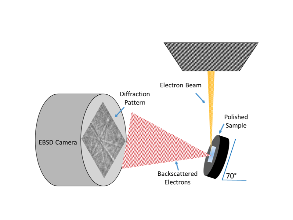
Electron backscatter diffraction (EBSD) microscopy is a characterization technique used with scanning electron microscopes (SEMs) for the determination of crystallographic information present in both metal and mineral samples. Much like energy dispersive X-ray spectroscopy (EDS), which provides the elemental composition through the interaction of electron beam and individual atoms, EBSD uses the interaction of the electron beam with the periodic arrangement of atoms in the (poly)crystalline region to generate diffraction (Kikuchi) patterns that can be captured with a unique camera-beam-detector geometry. The technique provides the ability to determine the crystalline phase and orientation information from the analyzed area in a bulk sample and supplements EDS data analysis when more detailed structural information is necessary.
EBSD serves as either a complement or alternative to other crystallographic analysis techniques such as transmission electron (TEM) microscopy and x-ray diffraction (XRD), with the added benefit of high-resolution, spatially resolved, phase/orientation mapping.
Unlike TEM analysis, where diffracted electrons pass through the analyzed region, EBSD imaging uses reflected electrons that have diffracted from atoms on the surface of the specimen. By operating as a surface analysis technique, EBSD is well suited for evaluating large polished surface areas, which is often infeasible with TEM, where extremely thin (nanometer) samples and consequently small areas are necessary to limit attenuation of the diffracted signal.
A frequent alternative to TEM is XRD, which uses the interaction of a (poly)crystalline sample with X-rays to produce Bragg diffraction that can be used to quantify the relative abundance of crystalline material phases that may be present in each specimen. Traditional XRD is well suited for the evaluation of powder samples, where randomized orientations yield better X-ray signals and the spatial distribution of phases in the powder solution is typically of minimal interest. Solid materials/polished samples can also be analyzed by XRD to yield texture information, but the analysis is typically used for the identification of preferred orientations via the generation of pole figures and is challenged in producing spatially localized texture information.
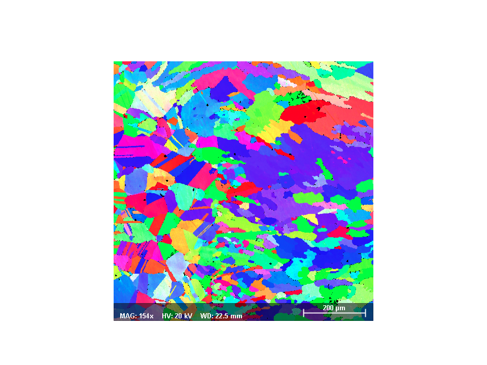
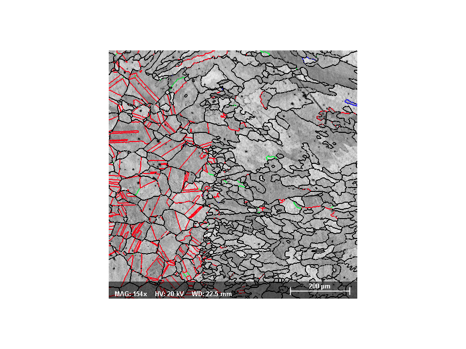
EBSD serves as a potential alternative to these two techniques, where the ability to scan and map large sample areas (mm2) is of interest. Grain size can be determined without the need for etching to reveal the location of grain boundaries1. Crystallographic phases can be evaluated when EDS information produces insufficient information as in the case of the iron oxides2 wüstite (FeO), magnetite (Fe3O4), and hematite (Fe2O3). Here, EDS is capable of identifying the presence of iron and oxygen but is limited in determining the atomic arrangement of these atoms.
EBSD can also be used to evaluate the crystallographic texture of a microstructure in the context of both grain and grain boundary orientations. Texture plays an important role in the macroscopic behavior of materials including corrosion behavior3, electro-magnetic properties4, and mechanical strength5.
The ability to map large areas of microstructure affords the production of texture metrics like pole figures, inverse pole figures, and orientation distributions. Grain boundary networks and grain boundary misorientation distributions can also be produced and assist in a greater understanding of the underlying microstructure and its relationship to material performance.
References
1ASTM E2627-13(2019), Standard Practice for Determining Average Grain Size Using Electron Backscatter Diffraction (EBSD) in Fully Recrystallized Polycrystalline Materials, ASTM International, West Conshohocken, PA, 2019
2Kim, Bae-Kyun, and Jerzy A. Szpunar. “Orientation imaging microscopy in research on high-temperature oxidation.” Electron Backscatter Diffraction in Materials Science. Springer, Boston, MA, 2009. 361-393.
3Dwivedi, Deepak, Kateřina Lepková, and Thomas Becker. “Carbon steel corrosion: a review of key surface properties and characterization methods.” RSC advances 7.8 (2017): 4580-4610.
4Bernier, Nicolas, et al. “EBSD study of angular deviations from the Goss component in grain-oriented electrical steels.” Micron 54 (2013): 43-51.
5Fizanne-Michel, C., et al. “Determination of hardness and elastic modulus inverse pole figures of a polycrystalline commercially pure titanium by coupling nanoindentation and EBSD techniques.” Materials Science and Engineering: A 613 (2014): 159-162.