


Contributors
Subscribe to our newsletter
Particles play a fundamental role in shaping our world and they can have a profound impact on society in ways that often go unnoticed. In order to control pollutants in the atmosphere, develop advanced pharmaceuticals, optimize food production processes, or create innovative materials for construction and electronics, we must be able to identify the size, shape and composition of particles.
 In fact, the study of particle sizes has become central to addressing some of the most pressing challenges within the fields of materials and environmental science as researchers work to prevent them from negatively impacting product quality, environmental sustainability, or human health.
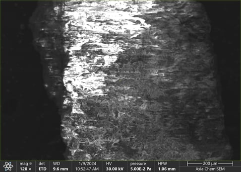
Researchers have a plethora of scientific technologies and techniques available to measure and characterize particles, including combining Scanning Electron Microscopy (SEM), to obtain detailed information about the surface morphology of a sample, with Energy Dispersive Spectroscopy (EDS) to obtain information about its elemental composition.
Identifying and classifying particles can even be automated using advanced software, such as IntelliSEM™ from RJ Lee Group, that controls the SEM/EDS to move and position samples, acquire images, map locations, and analyze data automatically.
While these amazing technologies increase efficiency and consistency, there are factors that must be considered. Before you choose any automation technology, you need to be aware of the most common pitfalls you could encounter, and how to overcome them.
Pitfall #1: Sample Preparation
Sample preparation for Scanning Electron Microscopy (SEM) is pivotal for acquiring high-resolution images, but it is fraught with potential pitfalls. For example, non-conductive specimens can accumulate charge when exposed to the electron beam, resulting in imaging artifacts and a lower throughput when automation software attempts to characterize charging artifacts.
To counteract this, samples may be coated with conductive materials provided they do not interfere with EDS analysis. Semi-conductive samples can also benefit from a coating with conductive metal (eg., palladium, gold, etc.), or by creating a conductive path using Al-tape or Cu-tape.
Another concern is contamination on the sample surface itself, which can reduce throughput of automated particle characterization and mask features of interest. To prevent this from happening, the material must be sufficiently rinsed and dried in a drying oven before plasma cleaning. The samples may even be stored in a desiccator to avoid moisture absorption. This applies only to samples that can withstand this type of cleaning, such as a polished metal surface for example. Training and iterative refinement of preparation techniques are essential to achieve optimal results.
Pitfall #2: Particle Size Range
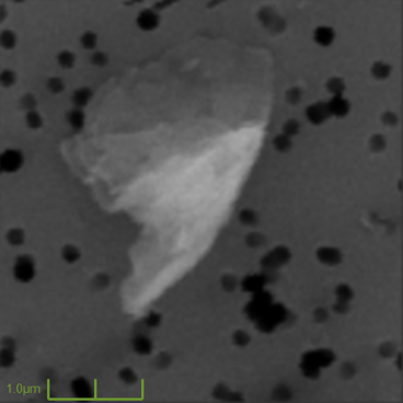
While the SEM has a large dynamic range in that it can see sub-micrometer detail as well as millimeter size features, a single pass is not ideal. At lower magnification levels, the electron beam moves more quickly across the sample, which may prevent the response signal from a small particle from reaching its maximum value and thus activating the detection threshold specified in the automation software.
When characterization of both small and large particulate is required, throughput and accuracy benefit greatly from a more deliberate approach. A commonly technique is to do an analysis of the full sample at a coarse magnification to characterize all large particulate.
Then, a second analysis of randomly selected sample areas is conducted at a higher magnification to characterize small particulate. This provides a good representation of the particulate population on the sample without the need to analyze the abundance of small particulate while allowing small particulate to be reliably detected.
Pitfall #3: Background Selection
Double-sided carbon tabs are one of the most popular mechanisms to mount particles for analysis with a SEM. However, as backscattered electrons are most often used to image particles of interest from the background, selecting a background with a sufficiently different average atomic number improves detection and sizing of particles.
There are a wide variety of media that can be used depending on the application, so for the best results, you should select an appropriate membrane filter from the options available including cellulose, polycarbonate, quartz, silver, or gold.
Pitfall #4: Securing particles
When imaging on a semiconductive background, particles can potentially ‘jump’ away when interacting with the electron beam, making results far less accurate. Securing particles by embedding them in carbon conductive tabs, coating with carbon or other conductive media, or embedding them in epoxy will helps to ensure reliable results.
Secured particles also allow for relocation should further analysis be required at a later time, provided that proper lab procedures are employed to prevent additional particulate deposits on the samples during storage.
Pitfall #5: Detector settings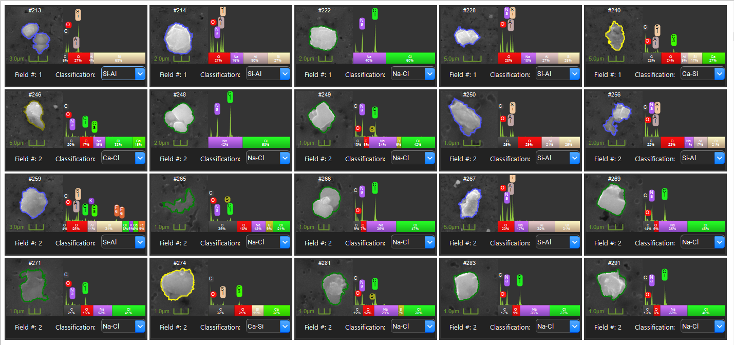
Researchers are often tempted to modify brightness and contrast settings based solely on the particles in a single image. For optimal repeatability, however, it is better to adjust these settings using multiple reference materials, such as Al-tape and Cu-tape. Additionally, when configuring brightness and contrast settings, remember to document any gamma correction applied to the imaging signal.
Summary
Particles are integral to numerous industries, influencing everything from environmental sustainability to pharmaceuticals to the quality of steel used in appliances and automobiles. As the significance of particulate becomes increasingly evident in our technologically advancing society, tools like SEM/EDS are vital for accurate characterization.
However, effective utilization of SEM/EDS requires meticulous attention to sample preparation, ensuring optimal particle size range representation, choosing appropriate backgrounds, securely anchoring particles, and consistent detector settings based on reference materials. Proper techniques and considerations are paramount for reliable and accurate particle analysis.